Using negative staining one can quickly preserve e.g. proteins, viruses and lipid vesicles to check their overall structure and concentration.
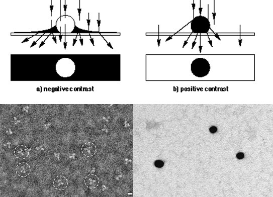
Negative staining is done by drying the sample on a carbon support film in the presence of a heavy metal salt solution, like e.g. Uranium, Lead, Molybdenum or Tungsten. The salts dry around the protein or virus particles and generate a reversed or negative contrast.
Negative staining is fast and easy to use specimen preparation technique that is commonly used as a first step in TEM. It is commonly used to identify particles in a suspension and have an initial view on what they look like.