Microscopy

The electron microscopy group houses several types of electron and light microscopes. We have different types of hardware and software implemented for correlative light and electron microscopy.


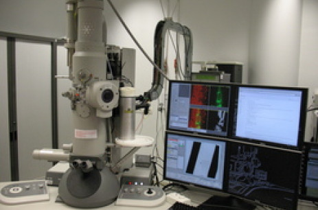
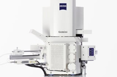
  • Zeiss gemini SEM 300

    The Zeiss Gemini SEM is a Field Emission Scanning Electron Microscope for imaging of micro structures. The microscope is equipped with the Gatan 3View system to image 3D ultrastructure using serial block-face imaging. Furthermore the system is also equipped with the Zeiss Atlas V system to allow imaging of serial sections in a non destructive manner. 

  • FEI Tecnai 20 FEG with iCorr

    The FEI Tecnai 20 FEG is a 200 kV TEM equipped with a Gatan 2001 energy filter with a 2k x 2K CCD camera, a Gatan US 4000 4k x 4K CCD camera and an FEI iCorr integrated light microscope for (cryo-) CLEM.

  • FEI T12 Spirit Biotwin

    The FEI Tecnai 12 BioTwin is a 120 kV TEM with a LaB6 filament and a FEI Eagle 4k x 4K CCD camera to record high contrast images. The microscope has in-house developed software for automated scanning of specimen grids, and is equipped for cryo electron microscopy and tomography.

  • FEi T12 Spirit Twin with iLEM and STEM

    The FEI Tecnai 12 Twin is a 120 kV TEM with a LaB6 filament and an FEI Eagle 4k x 4K CCD camera and is equipped with a integrated laser scanning microscope (iLEM) for correlative light-electron cryo microscopy and a Fischione HAADF STEM detector. Like the other TEMS this microscope runs in-house developed software for automated scanning of specimen grids and can perform cryo electron microscopy and tomography.

  • Zeiss Axio Imager with Linkam Freezing Stage

    The Zeiss Axio Image M2 is equipped with the Linkam CMS196M cryo correlative microscopy stage, which is controlled by Zeiss Zen software. It is specifically setup for automated cryo-CLEM imaging. It is capable of performing automated multi-color, z-stack recording and recording tile-scan overviews.

  • Leica DM RXA with Linkam THMS600 cryostage

    The Leica DM RXA is a development microscope, specifically used to develop super-resolution light microscopy on cryosamples. To that end, it is equipped with a Linkam THMS 600 freezing stage, 100x dry objectives (NA of 0.75 and 0.9), three laser lines (405, 488 and 532 nm) and a sensitive CMOS camera (PCO Edge 4.2). Acquisition and read-out is being done with home-written software specifically designed for super-resolution image acquisition.

Collaborate with us

Please contact us if you are interested in a scientific collaboration, electron microscopy services, or would like to be trained. 

Read more